Procedure: Cardiac Output Measurements

Ensure that patient and health care provider safety standards are met during this procedure including:

  • Risk assessment and appropriate PPE
  • 4 Moments of Hand Hygiene
  • Procedural Safety Pause is performed
  • Two patient identification
  • Safe patient handling practices
  • Biomedical waste disposal policies
  1. Prepare Monitor
  2. Connect Equipment and Review Setup
  3. Evaluate Proximal Port
  4. Confirm PA Waveform
  5. Prepare for Measurement


Checklist for Cardiac Output Measurement

Cardiac Output (CO) measurements are done in CCTC using the closed system CO-Set. Room temperature injectate (D5W 18-25 C) is the standard method.

Measurement of CO with a pulmonary artery catheter is done using the thermodilution technique:

  • A known volume of D5W at a temperature that is colder than blood temperature is injected into the right atrial port of the catheter. D5W is used because the molecular weight is incorporated into the cardiac output algorithm.
  • Cardiac output measurements are performed using the thermodilution technique. A known volume of blood (10 mL), at a known temperature (room temperature), is injected into the right atrium. Blood temperature is measured at the distal end of the pulmonary artery catheter and injectate temperature is measured as it enters the right atrial lumen. Cardiac output is determined by the change in blood temperature over time.
  • The syringe volume, injectate temperature and catheter size is programmed into the bedside computer by entering a computation constant.
  • Both PaceportTM and VIPTM catheter models use the same computation constant. This is automatically adjusted based on the injectate temperature.

Equipment Required

  1. Patient with a patent right heart catheter in good position (verified on x-ray by physician).               
  2. Cardiac output module (orange)
  3. Cardiac output cable with injectate and blood temperature measurement cables
  4. Closed-system CO Injectate Set with double barreled syringe and injectate thermistor
  5. 500 mL bag of D5W
  6. PPE - Non-sterile gloves
  7. Syringes and blood tubes for arterial and venous blood gases, lactate and hemoglobin as required.

Note: There are kits in plastic bags in the locked cupboard in the Bay 1 supply room. Each kit contains a double transducer set (one with blue stopcocks and one with yellow) for direct pressure connection to the blue (right atrial) and yellow (PA) ports. They also contain a cardiac output module and cardiac output cable with injectate temperature probe, cardiac output CO set with syringe).

PROCEDURE

1.

Prepare Monitor

  • Enter the patient's height and weight into the bedside monitor (BSA will automatically be calculated, which is required to confirm measured and calculated variables into indexed values (e.g., Cardiac Index, SVRI).
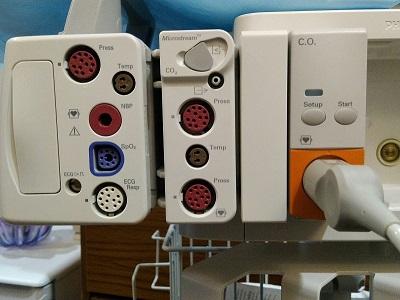
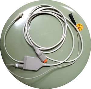
  • Plug in cardiac output module (Figure 1). Ensure the cable has two cables - one to connect to the pulmonary artery catheter and one to connect to the injectate port (Figure 2).
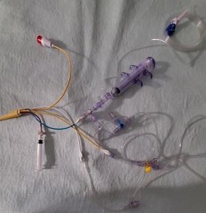
  • The pressure tubing with blue stopcock is connected to the blue port and the pressure tubing with the yellow stopcock is connected to the yellow port (Figure 3). 
  • Connect the cardiac output cable to the thermistor (white temperature connection) of the pulmonary artery catheter (Figure 4). Note that there are only 3 pins that connect into 4 receptacles within the thermistor( (Figure 5).


CO Module


CO Cables


Pressure Lines
Figure 1: Cardiac Output ModuleFigure 2: Cardiac Output CablesFigure 3: CVP and PAP Pressure Tubing

 2.

Connect CO Set

  • The right atrial and pulmonary artery lumens should already be individually connected to pressure monitoring (each lumen is continuously monitored with waveform displayed on monitor.
  • Confirm D5W as the injectate solution.
  • Prime the CO set tubing and syringe.
  • Connect ONE stopcock directly to the blue injectate lumen.  Connect the syringe to the stopcock so that the injection will be in a straight line. Additional stopcocks will increase the dead-space and reduce the injectate volume.
  • Leave the 6 inch extension tubing on the end of the pressure tubing and connect it directly to the stopcock without a needleless access device. Connect the pressure tubing at 90 degrees to the syringe flow.
  • Use the sampling stopcock near the end of the pressure tubing for bloodwork if needed. The right atrial lumen should display a continuous waveform.
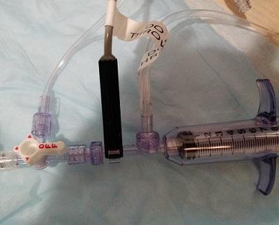
  • Connect the injectate probe clip to the syringe (Figure 3).  
  • Double check to confirm that the cardiac output cable is connected to the pulmonary artery thermistor.


Injectate Temperature


Injectate Clip Connected
Figure 4: Connect Injectate Temperature ClipFigure 5: Injectate Temperature Connected

 3.

Evaluate Proximal Injectate Port

Review proximal injectate infusions (blue port) and ensure there are no continuous infusions of vasoactive drugs prior to measuring cardiac outputs.

VASOACTIVE INFUSIONS SHOULD BE ADMINISTERED THROUGH THE INTRODUCER. THIS WILL ENSURE THEY REMAIN UNINTERRUPTED IF THE GANZ IS REPOSITIONED OR REMOVED.  

Continuous infusions of vasopressors/inotropes should be infused via the introducer. This prevents error during cardiac output measurements, and also ensures uninterrupted therapy even if the PA catheter is removed or repositioned.  If vasopressors are infused in either the PA injection or PA infusion port during repositioning, they may infuse extravascularly into the sleeve. 

Ensure that there are no continuous infusions (including insulin or heparin) infusing into the blue proximal injectate port of the pulmonary catheter.  If continuous infusions are being administered through the blue port, DO NOT perform cardiac output measurements because of the following:

  1. During cardiac output measurement, administration of the D5W injection will administer a bolus of any medication.
  2. During the period when cardiac outputs are being measured, the patient would be without their vasoactive medication.
  3. Upon completion of the cardiac output, the patient will be without the medication until the volume in the lumen has been reprimed with drug.
  4. Disruption will compromise the patient's cardiac output, and produce a cardiac output measurement that is invalid.

Assess infusions via introducer and proximal infusion port (VIP). Very rapid proximal infusions (> 100 ml/hr) or infusions of cold blood products may influence cardiac output measurements by changing the blood temperature.

 4.

Confirm PAP Waveform

Evaluate PA waveform and confirm pulmonary artery tracing to ensure proper catheter placement for cardiac output accuracy.

5.

Prepare for Measurement

Re-level transducer and obtain hemodynamic data including CVP. Collect data before performing cardiac output measurements for consistency. Contact Educator or CNS for PWP measurement if indicated; RNs should not routinely measure PWP due to infrequent performance of this skill.

Patient should be supine (head of bed can be elevated).

Post a paper recording of the CVP and PWP tracings.

6.

Measure Cardiac Output

  1. Unclamp D5W line of CO Injectate Set
  2. Slowly draw exactly 10 ml into syringe and close clamp (if you pull the plunger too hard the barrel of syringe will actually come apart)
  3. Turn stopcock toward pressure tubing and suspend IV infusion pump if a maintenance line is infusing.
  4.  maintenance IV to "off" and suspend infusion pump alarm.
  5. Press "Start CO" and wait until "Inject Now" is displayed (Figure 6, 7).
  6. Before injecting, identify where the injectate temperature will display. It is important to watch this temperature during the initial injection to ensure that the temperature drops. This provides confirmation that the injectate temperature is being correctly measured.
  7. Wait until the patient inspires and then inject the entire 10 ml using a steady and continuous technique. This will result in the injectate volume reaching the heart as the patient begins to exhale.
  8. Repeat at least 3 times, or until you have 3 acceptable CO curves with results within +/- 10%.  Touch the curve that you want to delete, touch again to restore (Figure 8).
  9. If height and weight has been entered, values will be indexed.
  10. Choose "calculate" to generate calculated values (e.g., SVRI, SVI) (Figure 9, 10,11).
  11. Print

NOTES: 

The double barrel syringe prevents premature warming of the injectate solution by the operator's hands.

The fall in the injectate temperature confirms that the injectate thermistor is correctly connected. A falsely low (often < 1.0) cardiac output can occur if there is a poor connection. Suspect if cardiac output is extremely low. When using room temperature solution, the greatest change occurs with the first injection when the luminal fluid is displaced for the first time.

Jerky injection technique will produce inaccurate readings as demonstrated by irregular cardiac output curves.

All injections should be delivered during the same phase of the respiratory cycle to avoid variable outputs due to breathing. The goal is end expiration when respiratory influence is most stable.

The timing of injection after the appearance of the "Inject Now" is not important. While older modules required injection within 4 seconds, current technology will wait for the change in the operator.


CO


Start CO


Edit CO
Figure 6:  Select Cardiac OutputFigure 7:  Select start Cardiac Output. Note where Tinj is located (middle right). Watch the Tinj during initial injection to confirm temperature change is being detected.Figure 8: Edit curves by touching the ones you want to exclude. Average update will be automatic.


HemoDynamic Calculations


Hemocalcs


Add data
Figure 9:  Select Cardiac OutputFigure 10:  Hemodynamics calculations displayed. Note that there is no PVR or PVRI calculation. This is because PWP has not been added (PVRI = PAP-PWP/CI *80)Figure 11:  To add data, touch the box with missing data and enter in the box that appears on the right.

7.

Resume Therapy

Turn stopcock to syringe to "off" and reclamp D5W line.

When final CO injection is done, resume right atrial pressure monitoring.  CVP and PA waveforms should be continuously displayed.  Set both pressures to a scale with a zero baseline to ensure prompt detection of right atrial or wedge positioning (do not choose optimize wave).  CVP should be be set to display systolic, diastolic and mean.

If the CVP diastolic and "PA" diastolic are close to the same and the "PA" systolic is higher than the CVP systolic, suspect right ventricular placement.

8.

Document

Record hemodynamic values in EHR. Enter the arterial and  mixed venous blood gas (drawn from the pulmonary artery catheter tip - called "SvO2") results into the oxygen-hemodynamic calculations and calculate. The SvO2 result is slightly different than the ScvO2.  

ScvO2 is drawn from an IJ or SC catheter. It only captures blood returning from the head and upper extremities.

SvO2 includes the oxygen that is remaining in the blood returning from the IVC, SVC and coronary sinus. This is a true "MIXED venous sample" as it captures all venous drainage, but prior to reoxygenation in the lung (only venous blood).

Prior to removal of a PA catheter it is useful to measure both a right atrial and pulmonary artery blood gas sample.  Measuring RA and PA blood before catheter removal enables subsequent interpretation of the RA blood. These two sample are not usually identical and following removal of the pulmonary artery catheter it can be difficult to know how to interpret (e.g., has the patient actually changed or the different results reflect the different sampling location)

Review results with physician.

Developed: August 3, 2006
Revised: February 4, 2021, Reviewed January 17, 2025
Brenda Morgan  MSc CNCC(C), CCTC

REFERENCES


1. Powers J, Chance R, Bortenschlager L, et al. Bedside placement of small-bowel feeding tubes in the intensive care unit. Critical Care Nurse. 2003; 23:16-24.

2. Lenart S, Polissar NL. Comparison of 2 methods for postpyloric placement of enteral feeding tubes. American Journal of Critical Care. 2003; 12:357-360.

3. Griffith DP, McNally AT, Battey CH, et al. Intravenous erythromycin facilitates bedside placement of post pyloric feeding tubes in critically ill adults: A double-blind, randomized placebo-controlled study. Critical Care Medicine. 2003; 31:39-44.

4. Booth, CM., Heyland, DK., Paterson, WG. (2002). Gastrointestinal promotility agents in critical care: A systematic review. Crit Care Med. 2002 Jul;30(7):1429-35.

5. Zaloga GP, Roberts PR. Bedside placement of enteral feeding tubes in the intensive care unit. Critical Care Medicine. 1998; 26:987-988.

6. Carroll GC. A Technique that improves the safety of feeding tube insertion. Critical Care Medicine. 2003; 31:1603-1604.

7. Thurlow PM. Techniques, materials and devices: Bedside enteral feeding tube placement in duodenum and jejunum. Journal of Parenteral Enteral Nutrition. 1986; 10:104-105.

8. Zaloga GP. Bedside method for placing small bowel feeding tubes in critically ill patients: A prospective study. Chest. 1991; 100:1643-1646.