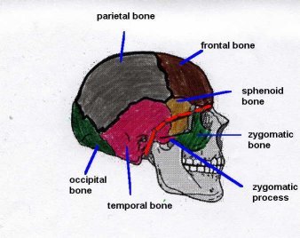
Where is the "Basal Skull"?
The skull bones surround the entire brain, extending underneath to create the base of the skull. The base of the skull is identified by the red line in Diagram 1. |
||
|
||
|
||
|
||
|
||
|
||
|
||
|
Diagram 3 |
||
|
-
Fossa Foramen Structures Contained in Foramen Function Signs/symptoms Anterior Fossa - cribiform plate
- CN I
- CN I - olfactory (ipsilateral sense of smell)
- anosmia (loss of smell)
- epistaxis (nose bleed)
- rhinorrhea (CSF from nose)
- optic foramen
- CN II (optic nerve)
- ophthalmic artery
- retinal artery
- CN II - optic (vision)
- visual loss or impairment
- impaired pupillary light response (CN II carries the light message to the CN III)
- periorbital hemorrhage (raccoon eyes)
- subconjunctival hemorrhage
- superior orbital fissure
- CN III
- CN IV
- CN V1
- CN VI
- CN III - oculomotor (ipsilateral up and down eye movement, eyelid opening, pupillary constriction)
- CN IV - trochlear (contra lateral downward and medial eye movement)
- CN V1 - 1st or ophthalmic division of the trigeminal nerve [V] (ipsilateral sensation of the cornea, nare and forehead)
- CN VI - abducens (ipsilateral movement of the eye in the temperal or lateral direction)
- impaired or dysconjugate eye movement
- ipsilateral ptosis (eyelid droop)
- ipsilateral pupillary dilation and loss of reaction
- loss of sensation to forehead, cornea or nare (loss of corneal reflex or nasal tickle response)
Middle Fossa - foramen rotundum
- CN V2
- CN V2 - 2nd or maxillary division of the trigeminal nerve [CN V] (ipsilateral sensation of the maxillary region of the face)
- loss of sensation to the mid face
- foramen ovale
- CN V3
- CN V3 - 3rd or mandibular division of the trigeminal nerve [CN V] (ipsilateral sensation of the mandibular region of the face)
- loss of sensation to the mid face
- ipsilateral weakness of masticator muscles
- foramen lacerum
- internal carotid artery
- sympathetic plexus
- supply of blood to anterior and middle cerebral cortex and ophthalmic artery
- cerebral cortex injury (upper motor neuron injury with contra lateral loss of motor function to face, upper and/or lower extremity; ipsilateral blindness)
- foramen spinosum
- middle meningeal artery and vein
- blood supply to temporal lobe
- temporal lobe injury (impaired hearing, comprehension, memory or seizure activity)
- epidural hematoma
- internal acoustic meatus
- CN VII
- CN VIII
- labyrinthine artery
- internal auditory artery
- CN VII - facial nerve - (ipsilateral facial movement, lacrimation, salivation, taste to anterior 2/3 of tongue, sensation around ear)
- CN VIII - vestibulocochlear nerve (hearing, balance)
- blood supply to labyrinth
- ipsilateral facial weakness
- ipsilateral inability to close the eye
- ipsilateral dry eye
- mouth dryness
- hemotympanium (blood in the ear canal)
- tinnitus
- hearing loss
Posterior Fossa - jugular foramen
- jugular vein
- sigmoid sinus
- CN IX
- CN X
- CN XI
- drainage of blood from brain
- CN IX - glossopharyngeal nerve (stimulates parotid gland, sensation to pharynx, soft palate, posterior third of tongue, auditory tube, tympanic cavity and carotid sinus)
- CN X - vagal nerve (muscles of soft palate and pharynx, parasympathetic control of heart and smooth muscles)
- CN XI - accessory (movement of neck and shoulders)
- echymosis behind the ear (battles sign)
- loss of gag reflex
- bradycardias
- inability to rotate neck
- hypoglossal canal
- CN XII
- CN XII - hypoglossal nerve (movement of tongue)
- inability to move tongue
- foramen magnum
- medulla oblongata
- meninges
- vertebral arteries
- meningeal branches of vertebral arteries
- spinal roots of CN XI
- medulla - respirations, blood pressure
- vertebral arteries - brainstem, occipital lobe and cerebellum
- bradypnea, respiratory irregularity
- hypertension and bradycardia
- cerebellar infarction (impaired balance or fine motor coordination)
- occipital lobe injury (loss of vision in the contra lateral visual field of both eye - e.g. right occipital lobe injury can cause loss of visual in the left field of the right and the left eyes)
References:
Barr, M., and Kiernan, J. (1993). The Human Nervous System: An Anatomical Viewpoint. (6th Edition). Lippincott: Philadelphia. pp 105-113, 122-147.
Diamond, M., Scheibel, A., & Elson, L. (1985). Human Brain Coloring Book. HarperPerennial: Toronto. pp 6-2.
Netter, F. (1997). Atlas of Human Anatomy. Novartis: New Jersey. pp 1-9.
Waxman, S. (1996). Correlative Neuroanatomy (23rd Edition). Appleton & Lange: Connecticut. pp 166-171.
Brenda Morgan, Clinical Nurse Specialist
November 19, 1999
Updated: January 19, 2019


